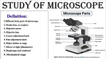
What is a microscope?
Use to magnify or magnify a micro-specimen to study the structure, size, and shape of cellular particles.
Definition:
It is a device that creates large images of small objects.
1 Different parts of the microscope body.
1. The ocular lens, or eyepiece
2. Objective Constellation
3. Objective lens
4. Coarse adjustment knob
5. Fine adjustment knob
6. Object holder or stage
7. Mirror or Light (Illuminator)
8. Diaphragm and Condenser
9. Mechanical phase
microscope types
What is a microscope?
Types Of Microscope:
There are different types of microscopes such as transmission electron microscopes (TEMs), scanning electron microscopes (SEMs), atomic force microscopes (AFM), near-field scanning optical microscopes (MSOM or SNOM, scanning near-field optical microscopy, and scanning tunneling microscopes (STM). )
However, the most common type of microscope is the optical microscope.
1. Electron microscope.
2. Light microscope
(It can be simple or compound).
A). Simple microscope:
A). Simple microscope:
This is a simple magnifying hand lens. Its magnification power is 2x to 200x.
b). Compound Microscope:
It has a lens battery, mounted in a.
complex device. It can be monocular or binocular.
1. A monocular consists of one eye piece.
2. Binoculars have 2 eye pieces.
Parts of Compound Microscope:
1. Stand gives stability.
2. Contains the body.
1) Body tube.
2) The stage
3) Knobs
1) Body Tube:
There are two tubes.
- Outer tube: houses the objective lens.
- Inner tube: houses the ocular lens.
2) Step:
glass slide.
It is a metal platform that gives space.
It has an aperture in the center that allows light to reach the object.
slightly below the stage could be a substage that
Consists of a condenser, through which light is focused onto an object.
3) KNOBS
The knobs are present on both sides of the body
"Adjustment" consists of two large knobs, movement under the body tube together with its lens.. "Fine Adjustment"
There are two small knobs on either side of the body.
3. Optical System:
Consists of eye pieces, objectives and condenser.
Eye piece:
Eyepiece magnification can be 5x, 10x, 15x.
- Objectives:
The magnification of objectives may be 4x, 10x, 40x, 100x.
- Condenser:
It is made up of two simple lenses and it condenses the light onto the object.
What is the function of a microscope?
A microscope is commonly used to study microscopic algae, fungi and biological samples.
What is magnification?
Magnification is that the method of maximising the relative size of Associate in Nursing object instead of its physical size. This growth is measured by a calculated variety known as "magnification".. Whenever this factor is much less than one, it corresponds to a reduction in size, also called minification or de-magnification.
What is resolution?
The term 'resolution' in research refers to the microscope's ability to tell apart the small print of Associate in Nursing object. In other words, it is the smallest distance at which two separate points on a specimen can still be seen as separate entities - either by an observer or a microscopic camera. The resolution of a microscope is related to the numerical aperture (NA) of the optical component as well as the wavelength of light used to examine the specimen.
What is depth of field?
Depth of field is defined as the space between the closest and farthest object planes that each can be in recognition at any given moment. In research, depth of field is however so much higher than and below the plane of the specimen the target lens and specimen will go. Go in perfect concentration.
What is an eyepiece?
The eyepiece, also known as the eyepiece, is the part used to see through a microscope. It is seen at the top of the microscope. Its customary magnification is 10x with Associate in Nursing elective lens that will increase magnification from 5X - 30X
What are objective lenses?
Objective lenses are large lenses used to view the specimen. Their magnification power is 40x-100x. A microscope has about 1-4 objective lenses mounted on it, some facing the refraction and some facing the front.
What is coarse adjustment?
A coarse adjustment knob moves the stage up and right down to bring the sample into focus.
What is fine adjustment?
The first-rate adjustment knob brings the specimen into sharp recognition underneath low energy and is used for all focusing whilst the use of excessive energy lenses.
What are Condenser Lenses?
Condensers are lenses used to collect and focus light from the illuminator onto the specimen. They are observed underneath the level subsequent to the diaphragm of the microscope. These play an important role in ensuring that clear sharp images are produced with high magnifications of 400X and above.
Safety Precautions for Microscope Use:
Lifting: Lift your microscope with two hands, one holding the arm or rear slot and the other supporting the base.
Table placement: Place the microscope on a flat, solid support that will not easily tip over. To prevent the cord from slipping, coil it.
Cleanliness: The lens must be clean to achieve resolution. Only lens paper or gauze and cleansing answer need to be used.. Do not remove any parts for cleaning; Doing so can cause dust to enter the microscope.


1 Comments
nice information
ReplyDeleteThank you